otseng wrote:
We see that the cilium is an example of a irreducible complex system.
Cilia are composed of at least a half dozen proteins: alpha-tubulin, beta-tubulin, dynein, nexin, spoke protein, and a central bridge protein. These combine to perform one task, ciliary motion, and all of these proteins must be present for the cilium to function. If the tubulins are absent, then there are no filaments to slide; if the dynein is missing, then the cilium remains rigid and motionless; if nexin or the other connecting proteins are missing, then the axoneme falls apart when the filaments slide.
What we see in the cilium, then, is not just profound complexity, but also irreducible complexity on the molecular scale. Recall that by "irreducible complexity" we mean an apparatus that requires several distinct components for the whole to work.
With such a lack of any viable molecular explanation for the origin of the cilium, it can be argued that it cannot have arisen from any naturalistic means. It must have been purposely designed.
This assumes that in order for a eukaryotic cell, any single-celled eukaryotic organism to survive at all, cilia is necessary. That is, there is no possibility that intermediate steps would be viable -- not even that there would be a natural-selection advantage that the lack of cilia provides, but that life would be viable at all.
This also assumes that these proteins could not operate as anything else inside cells, or even exist at all except as they exist in the axoneme. I reason this way because evolution is all about developmental mistakes that benefit organisms. It should be clear that when existing components are organized in different ways, different structures can be created. The irreducible complexity argument would therefore fall apart if we were to find these components operating in different ways inside a cell.
1) My
previous example of the centriole, which contains much of the building blocks & part of the structure of cilia
inside the cell.
2) To say that "removing" one of the components listed above would lead to a useless appendage is terribly misleading, because it implies that these components all came together at the same time to create the structure. For example, dyneins appear elsewhere in cells. During mitosis, dyneins and kinesins, as microtubule motors, assist with cetrosomal movement towards the poles of the splitting cell, which has nothing to do with flagella. Since they are present in other structures in the cell, this tends to support the hypothesis that cilia and flagella arose
from non-motile cells.
3) Not every single organism has the same structure of cilia & flagella:
Although the 9 + 2 pattern is the fundamental pattern of virtually all cilia and flagella, the axonemes of certain protozoans and some insect sperm show some interesting variations. The simplest such axoneme, containing three doublet microtubules and no central singlets (3 + 0) is found in Daplius, a parasitic protozoan. Its flagellum beats slowly (1.5 beats/s) in a helical pattern. Other axonemes consist of 6 + 0 or 9 + 0 arrangements of microtubules. These atypical cilia and flagella, which are all motile, show that the central pair of singlet microtubules is not necessary for axonemal beating and that fewer than nine outer doublets can sustain motility, but at a lower frequency.
Cilia and Flagella: Structure and Movement
This would tend to suggest that these structures did not necessarily arise spontaneously intact in the historical account, but that there may have been intermediate forms that provided "lower" levels of function. That they are different and viable suggests this.
4) The tubulins, the components of the microtubules that slide against each other to provide movements, are present in the microtubules inside the cell in the centromeres as well as those inside cilia (axonemes). Because both plant and animal cells have microtubules, but only animal cells have centromeres, we can infer that microtubules are not exclusively formed for cilia & flagella.
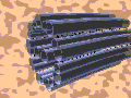
5) Here is a cross section of a cilia:

The
nexin bridge ("interdoublet nexin connection") is (theorized to be) the connection between microtubule doublets, acting like bungee cords so they don't slide past each other too much.
The microtubules in each doublet are not the same, they are distinguished as "A" and "B" tubules, as your quote points out. But there are also single and triplet microtubule structures that include "C" tubules, essentially the same as B's. Inside centrioles there are still other types: D, E, Z & H.
(
http://www.rpi.edu/dept/bcbp/molbiochem ... crotub.htm)
This suggests that the structure of the cilia was not, in fact, created out of thin air, but was already a potential possibility in prokaryotic cells.
6) Prokaryotes, in evolutionary terms older than eukaryotes, possess similar proteins, FtsZ, to those that make up tubules in eukaryotes:
A relatively new and interesting finding is that FtsZ and tubulin appear to be homologues.... it was also found that [Ftsz] could assemble into protofilaments, two-dimensional sheets, and protofilament rings in in vitrostudies, which was consistent with FtsZ having a cytoskeletal-like function.
Exploring structure and function of FtsZ, a prokaryotic cell division protein and tubulin-homologue
In short, these two proteins do the same things in different classes of cells. This article goes on to say, in technical detail, that these two proteins share sequences in important structures, such as what are called the "N-terminal" and the "C-terminal", both of which have to do with ATP-like protein binding and destruction. The article also goes on to say that these similarities could possibly be formed independently -- and they do not share all traits -- but it is more likely that they are related because of the core similarities they share. And, further, FtsZ is a strong candidate for an early evolutionary version of tubulins.